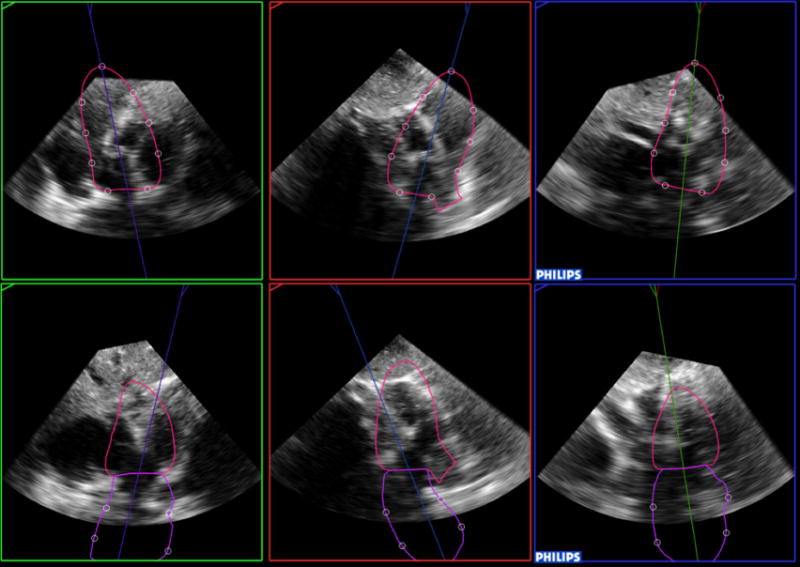
Figure 2.

Example of a failed analysis, both for the left ventricle and left atrium (top and bottom, respectively). The software was not able to correctly identify cardiac chambers and extracted from the 3D data set incorrect apical views, one of which (top left) coincidentally looks similar to a subcostal view.
