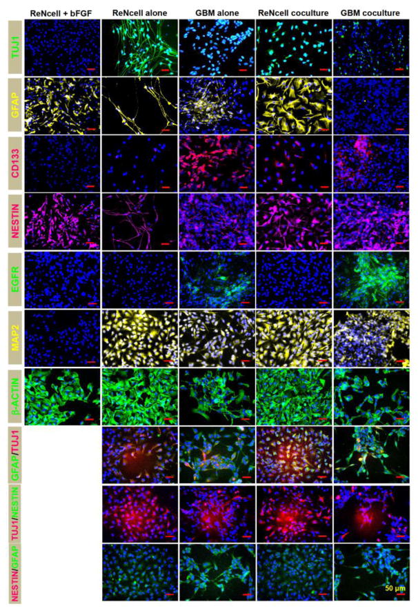
Figure 3.
Representative immunofluorescence images of ReNcells and GBMs, in standalone and cocultures, on day 10. For comparison, in select cases, ReNcells cultures in the presence of bFGF were also stained and imaged after 24 h in culture. Cultures were counterstained with DAPI for cell identification. Primary antibodies for TUJ1, GFAP, Nestin, CD133, EGFR, MAP2, and β-actin were used, with appropriate secondary antibodies. Double-immunolabeling was performed under select conditions. Scale bar: 50 μm.

