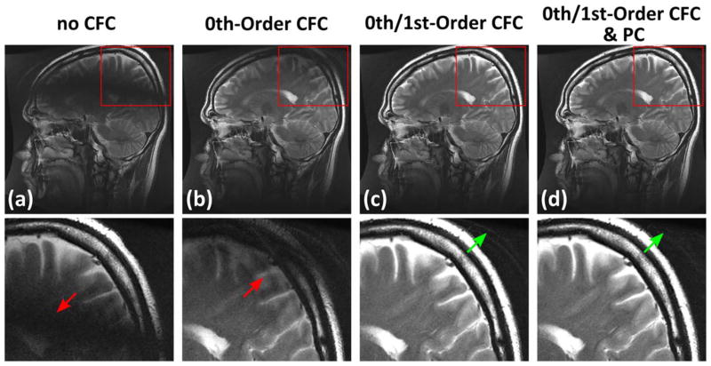
FIG. 6.
Examples of 2D sagittal T2 FSE images acquired on a healthy volunteer: a: image before CFC; b: with zeroth-order CFC; c: with zeroth and first-order CFC; d: with zeroth and first-order CFC as well as FSE PC. Note the black band caused by the first-order CF (red arrows). These effects are largely removed after CFC (c). The FSE PC applied after CFC can further suppress the ghosting (d) observed at the peripheral of FOV (green arrows).

