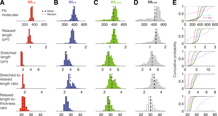
Figure 6.
Fibronectin with multiple Type III domain binding sites yield robust fibrillogenesis. Histograms for (row 1) the number FN molecules, (row 2) relaxed length, (row 3) stretched length, (row 4) stretched-to-relaxed length ratio, and (row 5) relaxed length-to-thickness ratio are shown for fibronectin with binding sites in (A, solid red in E) III1–3; (B, solid blue) III1–9; (C, solid green) III1–3,11; and (D, solid black) III1–15. Cumulative probability distributions are shown in (E), with distributions for fibronectin with single binding sites in III2 (dashed cyan) and III11 (dashed magenta) shown for comparison. Histograms are from 500 simulations (for A–C) and 1000 simulations (for D).

