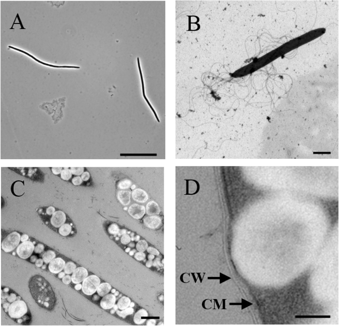
Figure 3.
Photomicrographs of strain AJ110941P grown in GAM medium under anaerobic conditions at 37 °C. (A) Phase-contrast photomicrograph. (B) Transmission electron micrograph of negatively stained cells. (C) Transmission electron micrograph of polyhydroxyalkanoate-like compounds accumulated in the cells. (D) Ultrathin section showing the Gram-positive cell wall (CW) and the cytoplasmic membrane (CM). Bars, 20 μm (A), 2 μm (B), 500 nm (C), and 100 nm (D).

