Figure 4.
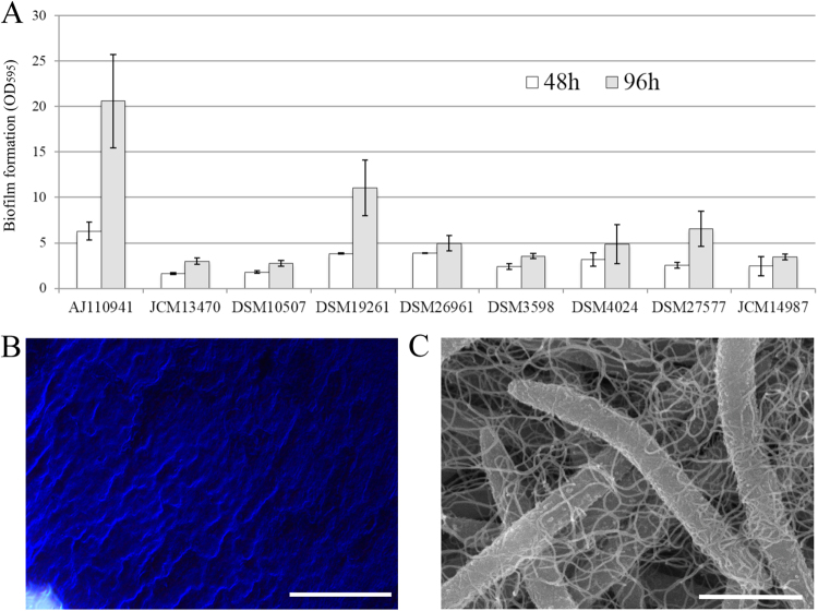
Comparison of biofilm forming-capacity among Lachnospiraceae strains. (A) Quantification of surface-attached biofilms of the Lachnospiraceae species grown in GAM medium at 37 °C for 48 h (white bars) and 96 h (gray bars), respectively. Biofilms were stained with crystal violet and quantified by measuring at 595 nm. The data represents the average of three biological replicates and the standard deviation is indicated by vertical bars. (B) Phase-contrast photomicrograph of biofilm-like aggregates of strain AJ110941P, after staining with crystal violet. (C) Scanning electron micrograph (SEM) of strain AJ110941P grown in GAM medium at 37 °C for 48 h. Bars, 20 μm (B) and 1.5 μm (C).

