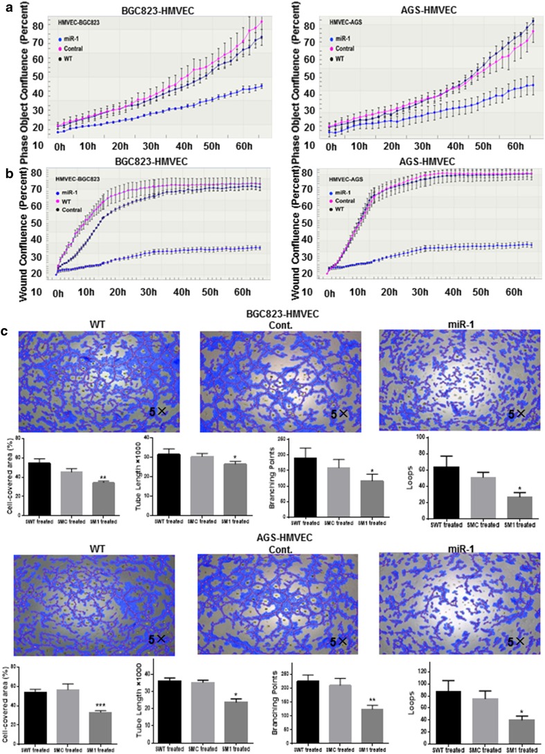
Fig. 5.
Effects of miR-1 on the proliferation, migration and tube formation of human microvascular endothelial cells. After both BGC823 and AGS cells had been transfected with GV268-miR-1 or GV268-ctrl plasmid respectively, the conditioned medium was collected and the effects of conditioned medium on human microvascular endothelial cell proliferation (a), migration (b), and tube formation (c) were assessed. The cell-covered area, tube length, branching points, and loops of tube formation were determined by WimTube from Wimasis Image Analysis (n = 3). One-way analysis of variance and Dunnett’s test were applied for analysis. Cont. control, HMVEC human microvascular endothelial cell, WT wild type, *P < 0.05, **P < 0.01, ***P < 0.001

