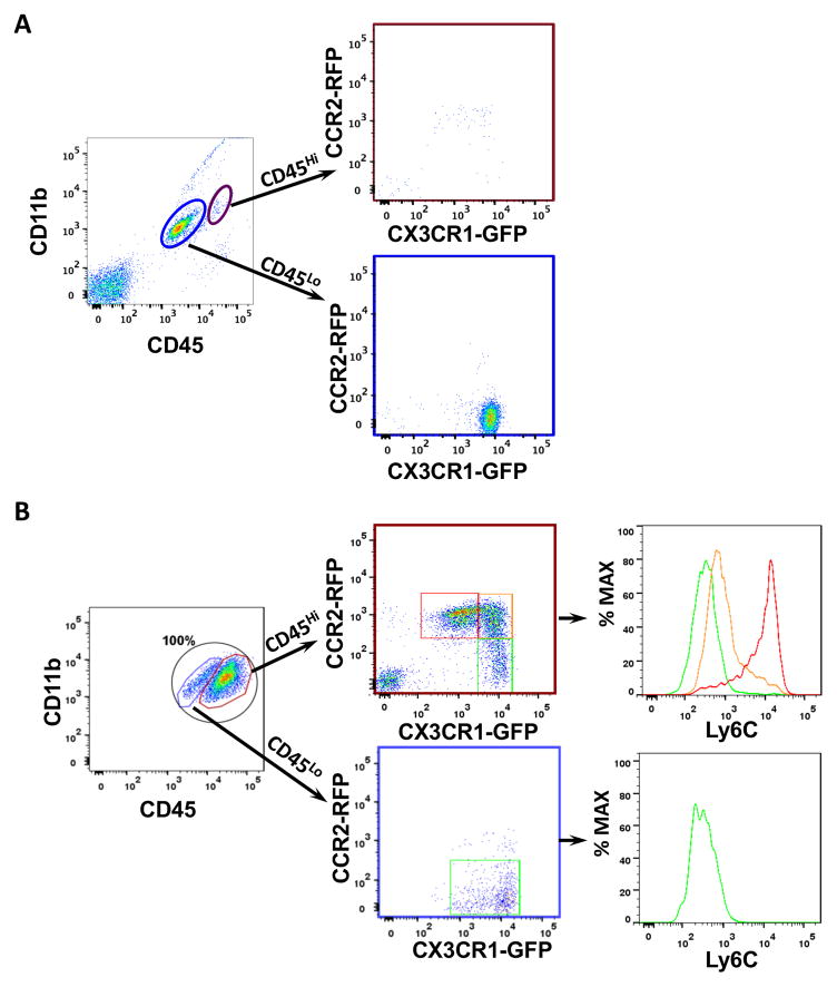
Figure 2. Cx3cr1GFP/WT;Ccr2RFP/WT knock-in mice distinguish resident microglia from BM-derived macrophages.
Representative dot plots gated on CD11b+CD45+ cells from (A) naïve and (B) tumors generated in Cx3cr1GFP/WT;Ccr2RFP/WT mice. (A) Magenta and blue circles delineate the CD11b+CD45Hi (blood-derived monocytes and macrophages) and CD11+CD45Lo (resident brain microglia) populations. Their CX3CR1-GFP and CCR2-RFP profiles are shown. (B) TAMs were identified by their CD11b and CD45 expression, and the CD45Hi and CD45Lo cells are further gated on RFP (CCR2) and GFP (CX3CR1) positivity. The CD45Hi population can be stratified into three related but distinct populations, whose Ly6C expression is examined. The CD45low population (microglia) expressed high level of CX3CR1-GFP, but little to no CCR2-RFP. Quantification of the subpopulations of CD45Hi or CD45Lo myeloid cells are described in the Result. N=3 for naïve mice and N=5 tumor bearing mice.

