
Fig. 5.
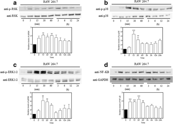
LPS-induced MAPK pathway activation. We performed a time-course experiment to evaluate the best times for the MAPK pathway activation. Macrophages were cultured with or without LPS (1 μg/mL) for 5, 15, 30, and 60 min and 3, 6, 12, and 24 h. Protein expression was evaluated using Western blot analysis. LPS induced a significant increase in the expression of (a) p-RSK, (b) p-p38, (c) p-ERK, and (d) NF-κB at different times. Data represent the mean ± SEM, n = 5. *p < 0.05, **p < 0.01, ***p < 0.001, versus sham