
Fig. 2.
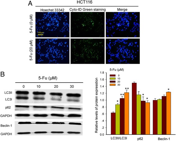
Immunofluorescent images of HCT116 cells treated with or without 5-Fu. a. Hoechst 33,342 staining (blue) indicates nucleus and Cyto-ID Green staining (green) autophagy status. b. Western blot analysis of beclin-1, p62 and LC3II/LC3I in HCT116 cells after exposing to varied concentrations of 5-Fu for 24 h. *, p < 0.05, and **, p < 0.01, compared to the vehicle (0 μM 5-Fu) cell group