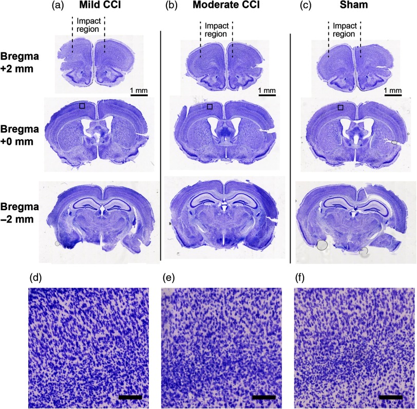
Fig. 7.
CV staining reveals negligible macroscopic anatomical damage 24 h after CCI. Columns (a) and (b) depict CV-stained coronal brain sections from mice that received “mild” and “moderate” CCI treatment, respectively, and (c) depicts CV-stained sections from sham experiments. Here, “mild” CCI corresponds to the parameter settings employed for all of the functional data in this study (velocity: ; dwell time: 100 ms; impact depth: 0.6 mm), while “moderate” corresponds to an increased impact depth (1.1 mm), with other parameters remaining the same. Slices from each condition are shown at locations on the AP axis corresponding to , 0 mm, and relative to bregma. Square subregions that are indicated in the slices at bregma (middle row) are shown on an expanded scale (imaged at , ) in (d)–(f).

