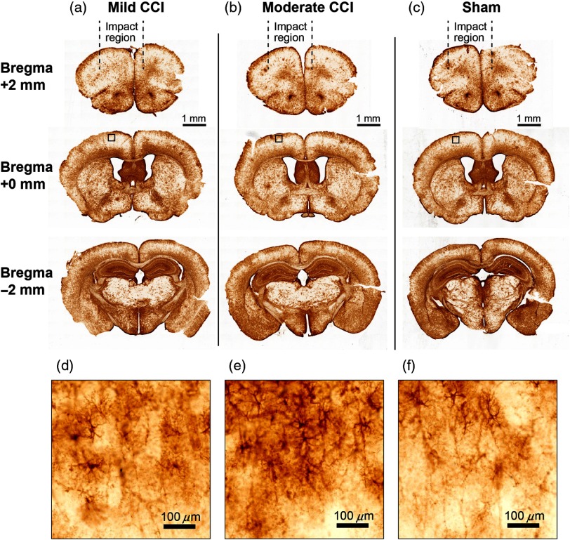
Fig. 8.
GFAP immunoreactivity 24 h after CCI indicates mild astrogliosis, but no macroscopic damage. Following the layout of Fig. 7, columns (a) and (b) depict GFAP immunoreactivity in coronal slices from mice that received mild and moderate CCI treatment, respectively, and (c) depicts results from sham experiments. Definitions of mild and moderate are as described in the caption for Fig. 7. Mild CCI corresponds to the parameter settings employed for all of the functional data in this study. The small notches on the ventrolateral corners of the brain slices indicate the hemisphere contralateral to the central axis of the impactor tip. Subregions that are indicated in the slices at bregma (middle row) are shown on an expanded scale in (d)–(f).

