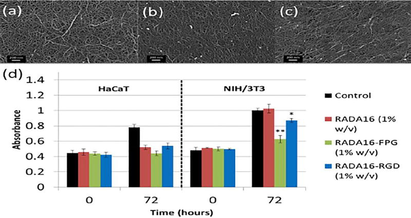
Figure 8.
a) Scanning electron microscopy of RADA16, (b) RADA16-FPG and (c) RADA16-RGD. (d) Cell viability result for the implemented keratinocyte (i.e. HaCaT) and fibroblast (i.e. NIH/3T3) using the three 1% w/v RADA16 NF scaffolds and cell control after 0 hour and 3 days of incubation. Reprinted from ref. [288], copyright 2014, Nature (licensed under a Creative Commons Attribution-NonCommercial-NoDerivs 4.0 International License).

