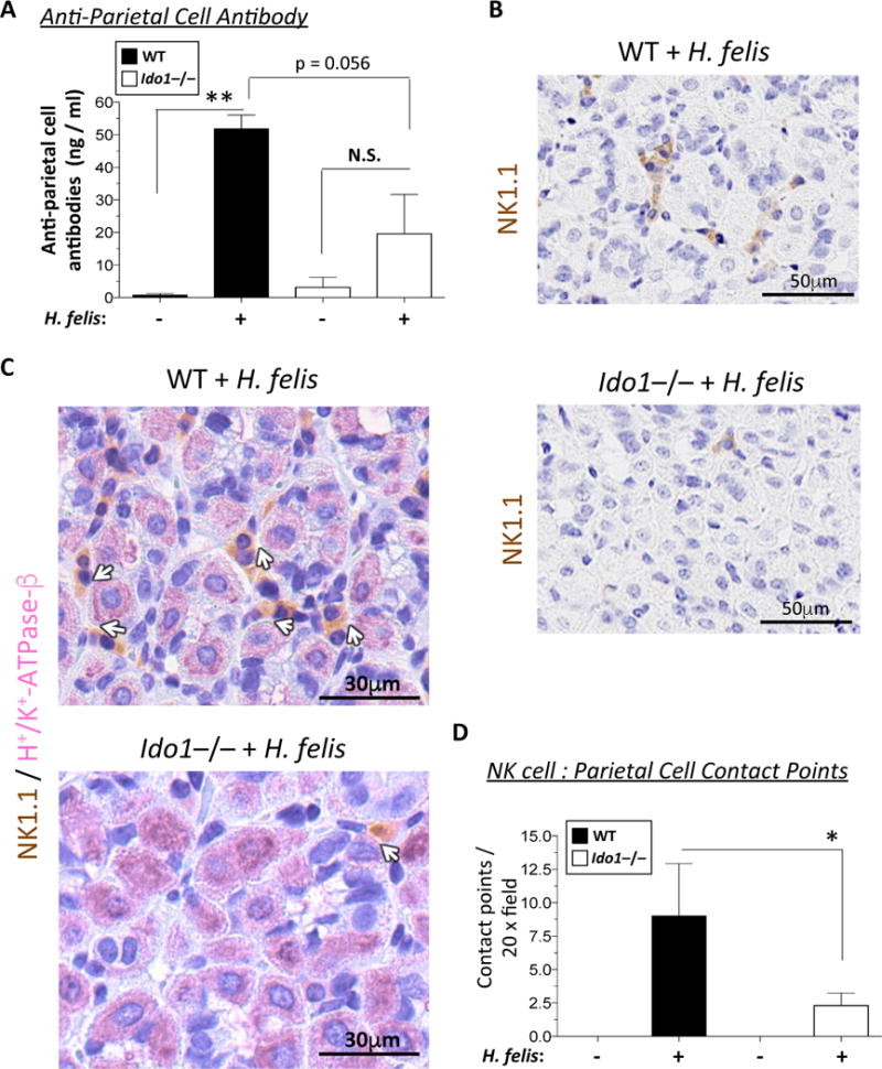
Figure 3. IDO1 deficiency associated with reduced autoantibody production and NK cell-to-cell contact with parietal cells.

A) Bar graph representation of plasma anti-parietal cell antibody levels in 6-month infected WT versus Ido1−/− mice compared to uninfected controls. B) Immunohistochemical staining of NK1.1-HRP antibody (brown) in 6-month infected WT versus Ido1−/− stomachs. These insets are obtained from the low power images shown in Suppl. Fig. 9A. C) Immunohistochemical double staining of NK1.1-HRP (brown) and H+/K+-ATPase-β (pink) of parietal cells in 6-month infected WT versus Ido1−/− stomachs. White arrows indicate the contact points between the NK cells and the parietal cells. D) Bar graph representation of morphometric quantification of NK cell/parietal cell contact points per 20x microscopic field. Data were compared using one-way analysis of variance (ANOVA) with Dunnet’s (parametric) or Dunn’s (non-parametric) for multiple comparison tests (Prism), Error bars represent the standard error of the mean. * P < 0.05. ** P < 0.01.
