Figure 1.

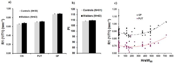
a) MRI longitudinal relaxation rates (R1) in basal ganglia regions of interest [caudate nucleus (CN), putamen (PUT), globus pallidus (GP)] for welders and controls; b) PI (pallidal index) for welders and controls. Data represent the mean ± standard errors (SEM).; c) R1 in the globus pallidus (GP) and putamen (PUT) as a function of welding hours (HrsW90) collapsed across welders and controls [modified from Lee et al. (2015)].
