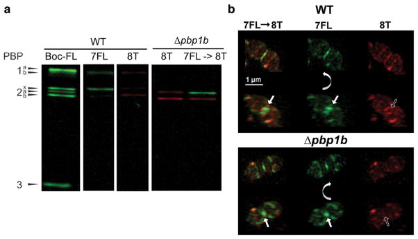
Figure 7.
Dual labeling with (2R,3S)-β-lactone-L-Phe-fluorescein (7FL) followed by (2R,3S)-β-lactone-D-Phe-TAMRA (8T). (a) This strategy enables separate visualization of the activity of PBP2x (green) and PBP2b (red) in gel-based analysis. Both probes were used at 5 μg/mL for 20 min. (b) Labeling with 7FL followed by 8T reveals distinct PBP localization patterns. Cultures of wild-type (IU1945) and Δpbp1b (E193) were grown, labeled with 7FL, washed, labeled with 8T, and imaged. Each set is two rows of images of the same cell, with the bottom row rotated relative to the top row around the indicated axis. In both strains, 8T labeling is excluded from the septum and restricted to peripheral rings when the cells are labeled first with 7FL. Solid arrows highlight central septal labeling, and empty arrows highlight the exclusion of 8T labeling from the center of the division site. These images are representative of ~40 mid-to-late division cells for each condition from two biological replicates.

