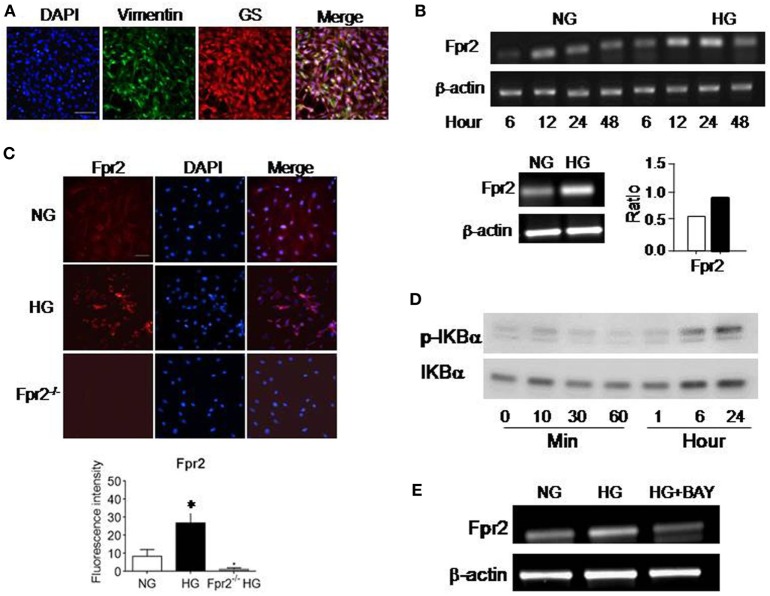
Figure 1.
The expression of formyl peptide receptor 2 (Fpr2) by mouse Müller glial cells (MGCs). Primary mouse MGCs were exposed to normal glucose (5.5 mM, NG) or high glucose (25.0 mM, HG) for 24 h. (A) Staining of the cells with vimentin (green) and glutamine synthetase (GS; red) to confirm the nature of MGC. (B) Increased Fpr2 mRNA in HG-treated MGCs. *Indicates significantly increased Fpr2 mRNA in HG-treated MGCs compared with cells treated with NG (p < 0.05). (C) Increased level of Fpr2 shown by fluorescence intensity in HG-treated MGCs. No Fpr2 immunoreactivity was detected in MGCs from Fpr2−/− mice. *Indicates significantly increased Fpr2 in fluorescence intensity in HG-treated MGCs compared with cells treated with NG (p < 0.05). (D) Western blotting showing phosphorylation of IκBα in MGCs induced by HG at the indicated time points. (E) The effect of IκB/NF-κB inhibitor BAY 11-7082 on Fpr2 expression by MGCs under HG for 24 h.

