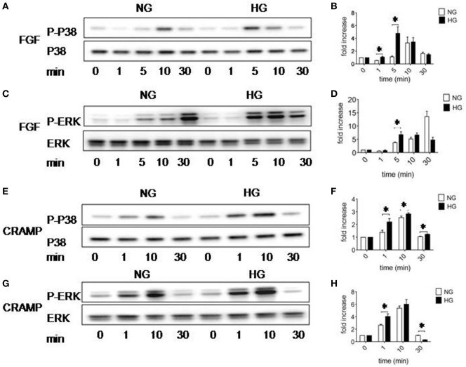
Figure 5.
Activation of p38 and ERK1/2 MAPK in Müller glial cells (MGCs). Western blotting was performed to examine the phosphorylation of p38 and ERK1/2 MAPKs in MGCs. (A) p38 phosphorylation induced by fibroblast growth factor (FGF) (10 ng/ml) in MGCs cultured with normal glucose (NG) or high glucose (HG). (B) Densitometry quantification of phosphorylation p38 (P-p38) normalized against total p38 based on results shown in (A). The results are presented as fold changes. *Indicates significantly (p < 0.05) increased FGF-induced P38 phosphorylation in MGCs cultured in HG compared to cells in NG. (C) ERK phosphorylation induced by FGF (10 ng/ml) in MGCs cultured with NG or HG. (D) Densitometry quantification of P-ERK normalized against total ERK. The results are presented as fold changes. *Indicates significantly (p < 0.05) increased ERK phosphorylation induced by FGF in MGCs cultured in HG compared to cells in NG. (E) p38 phosphorylation induced by cathelin-related antimicrobial peptide (CRAMP) (10−6 M) in MGC cultured with NG or HG. (F) Densitometry quantification of P-p38 normalized against total p38. The results are presented as fold changes. *Indicates significantly (p < 0.05) increased p38 phosphorylation induced by CRAMP in MGCs cultured in HG compared to cells in NG. (G) ERK phosphorylation induced by CRAMP (10−6 M) in MGCs cultured with NG or HG. (H) Densitometry quantification of P-ERK normalized against total ERK. The results are presented as fold changes. *Indicates significantly (p < 0.05) increased ERK phosphorylation induced by CRAMP in MGCs cultured in HG compared to cells in NG.

