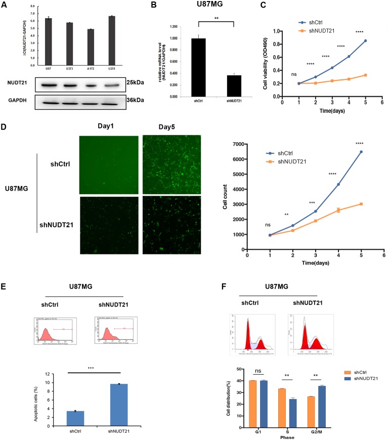
FIGURE 2.
Knocking down NUDT21 suppresses glioblastoma cell proliferation. (A) The results of RT-qPCR indicated that all four glioblastoma cell lines could be of high-expression of NUDT21 [ΔCt = Ct value of NUDT21 – Ct value of GAPDH (control gene), mean ± SD, n = 3]. The U87MG and U251 cell lines were selected for further study to investigate the biological role of NUDT21 in GBM cells. (B) NUDT21 knockdown was accomplished using shRNAs in glioma cells (mean ± SD, n = 3, ∗∗p < 0.01). (C) MTT assay showed that, compared with the cell proliferation in the control cells, NUDT21 knockdown significantly decreased cell proliferation in U87MG cells. The OD values were measured at indicated days (mean ± SD, n = 3, ns, significant; ∗∗∗∗p < 0.0001). (D) Celigo assay was performed to detect the cell viability over 5 days after transfection. The colony numbers of U87MG cells transfected with shCtrl were evidently higher than those transfected with shNUDT21 (mean ± SD, n = 3, ns, significant; ∗∗p < 0.01; ∗∗∗p < 0.001; ∗∗∗∗p < 0.0001). (E) To further determine the physiological role of NUDT21 in cell growth, U87MG cells were transfected with shNUDT21. After 48 h, the apoptotic rates of cells were detected by flow cytometry (mean ± SD, n = 3, ∗∗∗p < 0.001). (F) Flow cytometry analysis was performed to further examine the effect of NUDT21 on the proliferation and altered cell cycle progression of GBM cells. The bar chart represented the percentage of cells in G0/G1, S, or G2/M phase (mean ± SD, n = 3, ns, significant; ∗∗p < 0.01 versus shCtrl group at indicated phase).

