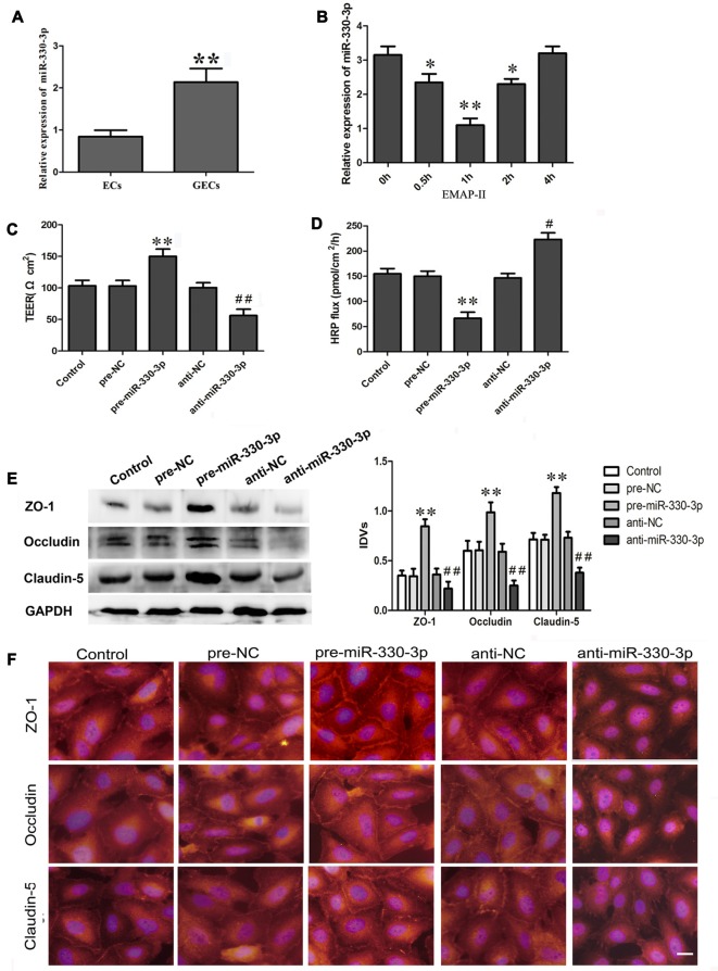
Figure 1.
(A) The endogenous expression of miR-330-3p in ECs and glioma microvascular endothelial cells (GECs). U6 was used as an inner control. Data represent means ± standard deviation (SD; n = 5, each). **P < 0.01 vs. ECs group. (B) Effect of Endothelial monocyte-activating polypeptide-II (EMAP-II) on the expression of miR-330-3p in GECs. U6 was used as an inner control. Data represent means ± SD (n = 5, each). *P < 0.05 and **P < 0.01 vs. EMAP-II 0 h group. (C,D) Transendothelial electric resistance (TEER) and horseradish peroxidase (HRP) assays were used to measure the effects of overexpression or silencing of miR-330-3p on the permeability of blood–tumor barrier (BTB). (E,F) The expression and distribution of tight junction (TJ) related proteins in GECs after overexpression or silencing of miR-330-3p. GAPDH was used as an inner control. Data represent means ± SD (n = 5, each). **P < 0.01 vs. pre-NC group; #P < 0.05 and ##P < 0.01 vs. anti-NC group.

