
Figure 2.
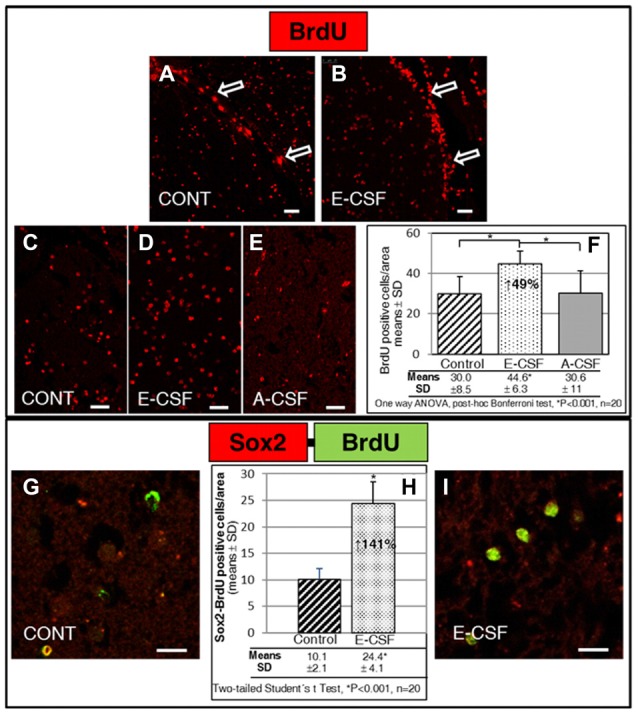
Effect of Embryonic CSF (E-CSF) on mitotic activity in SVZ niche NSCs, monitored by nuclear incorporation of BrdU (red). Confocal photomicrographs (A,B show the SVZ of “in vitro” cultured adult mice brain slices (see Figure 1B). Note the substantial increase in NSCs mitotic activity under the ventricular surface (white arrows) induced by E-CSF (B), compared with the controls (A). Confocal photomicrographs (C–E) show the Striatum area of “in vitro” cultured adult mice brain slices close to the SVZ (ST in Figure 1B). Quantification of BrdU positive nuclei in Control (C), E-CSF (D) and A-CSF (E) treated brain slices was plotted in graph bars (F) and expressed as means ± SD (n = 20); the significant threshold was set at p ≤ 0.001 (*) according to the one-way ANOVA, post hoc Bonferroni test. The results reveal a statistically significant increase (49%) in the number of BrdU positive NSCs in CSF-E treated brain slices with respect to the controls and A-CSF treated slices; this suggests a specific activation by E-CSF of mitotic activity in NSCs of the SVZ niche. Confocal photomicrographs (G,I) correspond to the striatum area of “in vitro” cultured adult mice brain slices, close to SVZ (ST in Figure 1B). Images show double immunolabeling with antiBrdU (green) and antiSox2 (red) antibody; co-localization of both antibodies was interpreted as NSCs dividing (BrdU positive) but not differentiating (Sox2 positive). Quantification of co-labeled BrdU and Sox2 NSCs in Control (G) and E-CSF treated brain slices (I) was plotted in graph bars (H) and expressed as means ± SD (n = 20); the significant threshold was set at p ≤ 0.001 (*) according to the two-tailed Student’s t-test. The results reveal a statistically significant increase (141%) in the number of NSCs that undergo replication but remain undifferentiated in CSF-E treated brain slices, in comparison with the controls. Scale bar: 25 μm (A–E) and 10 μm (G,I).