
Figure 3.
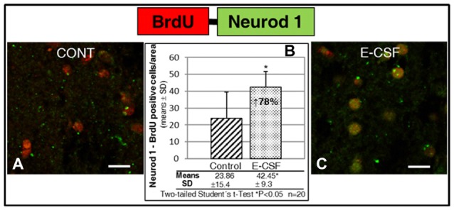
Effect of E-CSF on the number of NSCs with neuronal commitment, monitored by co-expression of BrdU (red) and Neurod1 (green). Confocal photomicrographs show the Striatum area of “in vitro” cultured adult mice brain slices close to the SVZ (ST in Figure 1B). Quantification of co-labeled BrdU and Neurod1 NSCs in Control (A) and E-CSF treated brain slices (C) was plotted in graph bars (B) and expressed as means ± SD (n = 20); the significant threshold was set at p ≤ 0.05 (*) according to the two-tailed Student’s t-test. The results reveal a statistically significant increase (78%) in the number of NSCs with neuronal commitment in CSF-E treated brain slices with respect to the controls; this suggests an increase in adult brain neurogenesis activity induced by embryonic CSF. Scale bar: 10 μm (A,C).