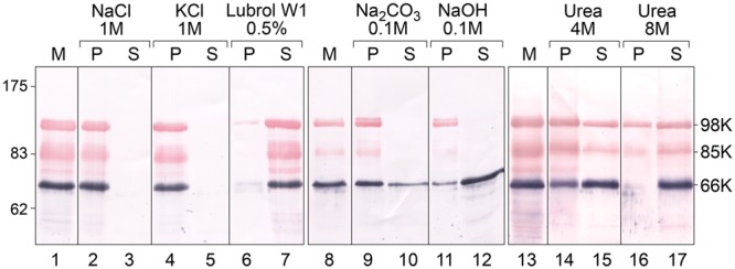
FIGURE 2.

Analysis of the association of TYMV replication proteins with membranes by ionic, alkaline and urea extraction. Membrane fractions (M) were obtained from TYMV-infected Chinese cabbage tissues and were incubated in medium containing 1 M NaCl (lanes 2–3); 1 M KCl (lanes 4–5); 0.5% Lubrol W1 (lanes 6–7); 0.1 M Na2CO3, pH 11 (lanes 9–10), 0.1 M NaOH (lanes 11–12), 4 M urea (lanes 14–15) or 8 M urea (lines 16–17). Soluble (S) and insoluble pellet (P) fractions were then separated by centrifugation, and each fraction was subjected to 8% SDS-PAGE and immunoblot analysis. Protein samples were revealed sequentially using anti-66K and anti-98K antisera and NBT/BCIP (purple) and Fast Red/Naphtol (red) substrates, respectively. Lanes 1–7, lanes 8–12, and lanes 13–17 correspond to different tissue samples processed and analyzed independently. Molecular mass markers (Biolabs) are indicated on the left, whereas positions of the viral proteins 98K, 85K, and 66K are indicated on the right.
