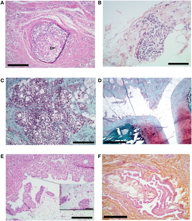
Figure 6.
Histological analysis of synovial proliferation. Sections with hematoxylin eosin (A,B,E,F) or safranin O and Fast Green (C,D) stain. (A) Synovial membrane of knee of an animal that received intra-articular (IA) injection containing incomplete Freund’s adjuvant (IFA). Arrowhead shows multinucleated gigantic cell. 10× magnification, bar 200 µm. (B) Synovial membrane of right knee of an animal that received IA boost with citrullinated peptides + IFA and presented prolonged swelling. 20× magnification, bar 100 µm. (C) Knee synovial membrane. 10× magnification, bar 200 µm. (D) Knee synovial membrane, bone, and cartilage of the same animal. 4× magnification, bar 500 µm. (E) Right knee synovial membrane of an animal that received an IA boost with citrullinated peptides + IFA and presented prolonged swelling. Insert shows fibrin and leukocyte in the synovial cleft. 10× magnification, bar: 200 µm, insert bar 50 µm. (F) Section of metacarpophalangeal joint synovial membrane of an animal without clinically identified manifestation on these joints. 10× magnification bar 200 µm.

