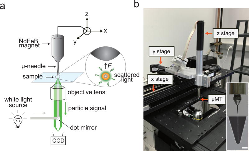
Figure 6. Dark-field microscope and micro-magnetic tweezer (µMT) setup.
(a) A schematic illustration of µMT and dark-field microscopy setup. Steel needle (tip radius: 5 µm) - NdFeB magnet (diameter 1 cm) assembly is mounted to the xyz translation stage. A 4 mm ellipsoidal dot mirror is installed at a filter cube set for a dark-field illumination. (b) A photograph of experimental setup. Scale bar, 20 µm.

