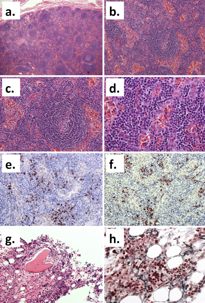
Figure 3.
The histological findings. (a-d) The histological appearance of the left axillary lymph node (Hematoxylin and Eosin (H&E) staining). (a) The basic structure of the lymph node is obscure (original magnification: ×40). (b) Many lymphoid follicles with unclear atrophic germinal centers and the expansion of the interfollicular zone are apparent (original magnification: ×100). (c) The peripheral mantle layer was developed with a concentric cellular distribution (original magnification: ×200). (d) Arborized blood vessels were present and the infiltration of small lymphocytes and plasma cells in the interfollicular zone was noted (original magnification: ×400). (e, f) Kappa (e) and Lambda (f) immunostaining showed no clear monoclonality (original magnification: ×200). (g, h) The histological appearance of the bone marrow. (g) An increase in the number of megakaryocytes was evident on H&E staining (original magnification: ×200). (h) Silver impregnation staining showed an increase in reticular fibers (original magnification: ×400).

