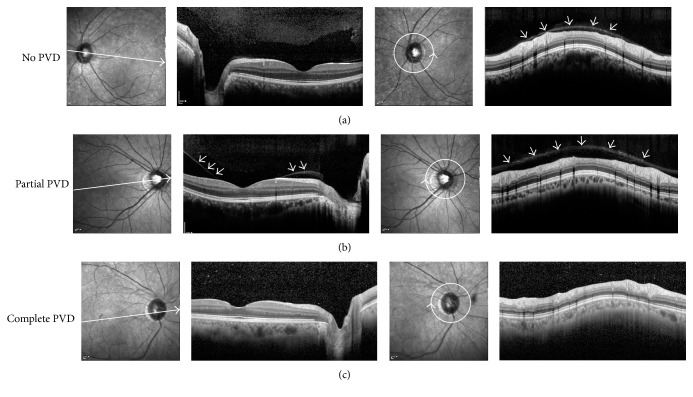
Figure 2.
Posterior vitreous detachment grading by optical coherence tomography. Posterior vitreous status was graded with optical coherence tomography. (a) No posterior vitreous detachment (PVD), with complete attachment of the posterior vitreous cortex (PVC) to the perifoveal area, fovea, and optic disc. Circumferential peripapillary scan also confirms attached PVC (arrows). (b) Partial PVD with separation of the PVC over perifoveal area (arrows). Circumferential peripapillary scan shows attached PVC (arrows). (c) Complete PVD with detachment of PVC over perifoveal area, fovea, and optic disc, showing optically empty space above. Circumferential peripapillary scan also lacks attached overlying PVC.

