Figure 2.
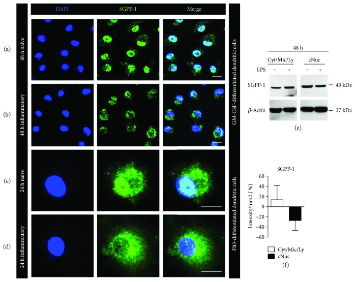
(a, b) Representative pictures of confocal laser microscopy of untreated mature GM-CSF-differentiated CD11c+ dendritic cells after 48 h of starvation (naive) and 48 h of LPS stimulation (inflammatory) stained with DAPI (blue) and anti-SGPP-1 (green) (n = 7). (c, d) Representative pictures of confocal laser microscopy of untreated mature Flt3-differentiated CD103+ dendritic cells after 24 h of starvation (naive) and 24 h of LPS stimulation (inflammatory) stained with DAPI (blue) and anti-SGPP-1 (green) (n = 2). (e) Representative Western blot stained with anti-SGPP-1 antibody and anti-β-actin antibody in crude cytosolic (Cyt/Mic/Ly; Cyt = cytosol, Mic = microsomes, and Ly = lysosomes) and crude nuclear (cNun) fraction of cell lysates of naive GM-CSF-differentiated dendritic cells or 48 h upon LPS stimulation (n = 3). (f) Quantification of Western blot bands of anti-SGPP-1 staining after stimulation with LPS.

