FIGURE 2.
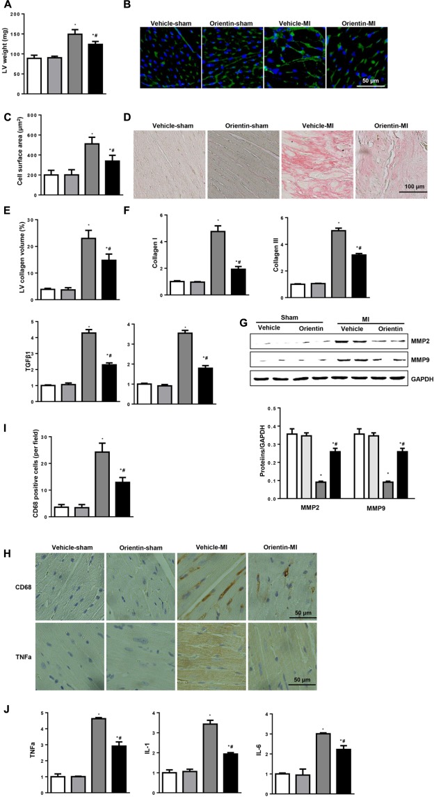
Orientin attenuates MI-induced fibrosis and inflammatory response. (A) LV weight in the indicated mice 4 weeks after MI (n = 6–8). (B) Immunofluorescent staining of wheat hemagglutinin (WGA) (n = 6). (C) Analysis of cell surface area (n > 200 cell per group). (D) PSR staining of heart to analyze the LV collagen volume (n = 25+ fields per experimental group). (E) Statistical analysis of the CSA and LV collagen volume (%). (F) The relative mRNA levels of collagen I, collagen III, TGFβ1, and CTGF, in LV samples from vehicle and orientin-treated mice heart (n = 6). (G) Representative Western blots and quantitation of MMP2, MMP9 in the heart tissue of vehicle and orientin-treated mice at 4 weeks after sham or MI surgery (n = 6). (H) Immunohistochemistry staining showing the number of CD68-positive cells and TNFα release in the heart cross-sections of the vehicle and orientin-treated mice. (I) Statistical analysis of the number of CD68-positive cells in the indicated group (n = 6). (J) The relative mRNA levels of TNFα, IL-1 and IL-6 in samples from vehicle and orientin-treated mice heart (n = 6); ∗p < 0.05 vs. vehicle-sham; #p < 0.05 vs. vehicle-MI.

