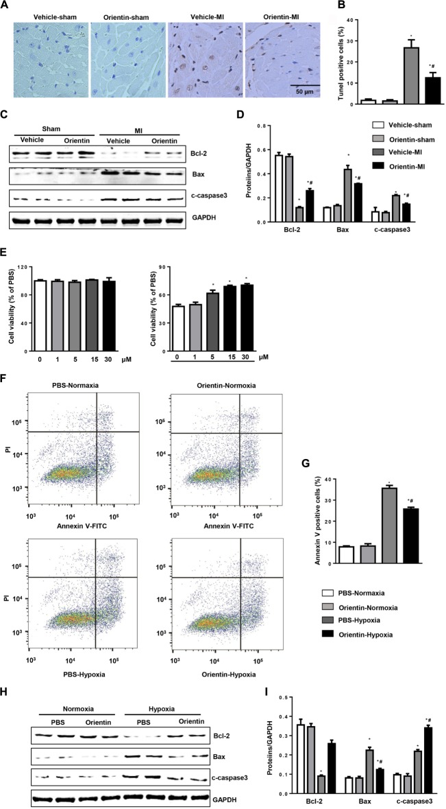
FIGURE 3.
Orientin inhibits MI-induced apoptosis. TUNEL staining (A) and quantitation (B) in the hearts of vehicle and orientin-treated mice at 4 weeks post-MI (n = 6). (C,D) Representative Western blots and quantitation of Bax, Bcl-2, and C-caspase3 in the heart tissue of vehicle and orientin-treated mice at 4 weeks after sham or MI surgery (n = 6); ∗p < 0.05 vs. vehicle-sham; #p < 0.05 vs. vehicle-MI. (E–I) NRCMs were pretreated with orientin (1, 5, 15, 30 μM) for 12 h, then exposed to hypoxia for 24 h. (E) MTT assays were performed to detect cell viability (n = 5, ∗p < 0.05 vs. hypoxia-0 μM group). Annexin V staining (F) and quantitation (G) in the indicated group (n = 6). Representative Western blots (H) and quantitation (I) of Bax, Bcl-2, and C-caspase3 in the indicated group (n = 6); ∗p < 0.05 vs. vehicle-normoxia; #p < 0.05 vs. vehicle-hypoxia.

