Figure 4.

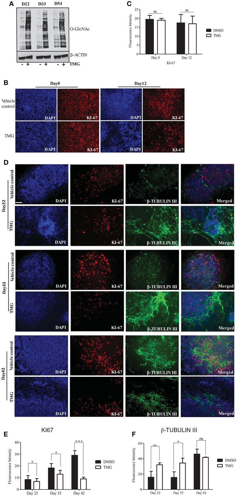
Higher O-GlcNAc levels affect neural progenitor proliferation and differentiation during corticogenesis. Neural progenitor cells from Day12 of neural induction were differentiated for cortical neurons in N2/B27 media for up to day 54. (A) Western blot analysis of TMG treated and vehicle control samples were performed from D22, D33, and D54 of cortical differentiation of neural progenitors to detect total O-GlcNAc levels. The expression of β-ACTIN was used as loading control. The data are representative of three independent biological replicates. (B) Immunostaining of TMG treated and vehicle control H9 cells for KI-67 at Day0 and Day12 of neural induction. (C) The fluorescence intensity of images from panel (B) was quantified using ImageJ (Fiji) software. The data are represented as mean ± standard deviation. (D) Immunostaining of cortical differentiated TMG treated and vehicle control cells for KI-67 and β-Tubulin III at Day22, Day33, and Day42 of differentiation. In (B,C), the cells were imaged with fluorescence microscope. DAPI represents nuclear staining. Scale bar = 100 μm.The data are representative of three independent biological replicates. (E,F) The fluorescence intensity of images from panel (D) was quantified using ImageJ (Fiji) software. The data are represented as mean ± standard deviation. *p ≤ 0.05, **p ≤ 0.01, ***p ≤ 0.001 (two-tailed unpaired Student's t-test).
