
Figure 2.
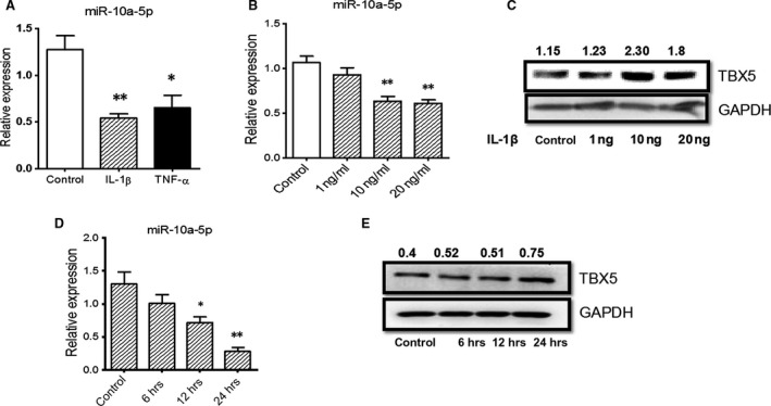
IL‐1β regulated the expression of miR‐10a‐5p and TBX5 in SW982 cells. SW982 cells were stimulated with IL‐1β and TNF‐α for 24 hrs, and relative expression of miR‐10a‐5p (A) was detected by RT‐qPCR. The cells were then stimulated with different doses of IL‐1β for 24 hrs (B and C) or with 10 ng/ml IL‐1β for different time‐points (D and E), respectively. miRNA expression was detected by RT‐qPCR, whereas TBX5 expression was determined by Western blotting. The expression levels of miR‐10a‐5p and TBX5 were normalized by U6 snRNA and GAPDH, respectively. The data represent the means ± S.E.M. of three independent experiments. Levels of significance were calculated using Student's t‐test (*P < 0.05, **P < 0.01).