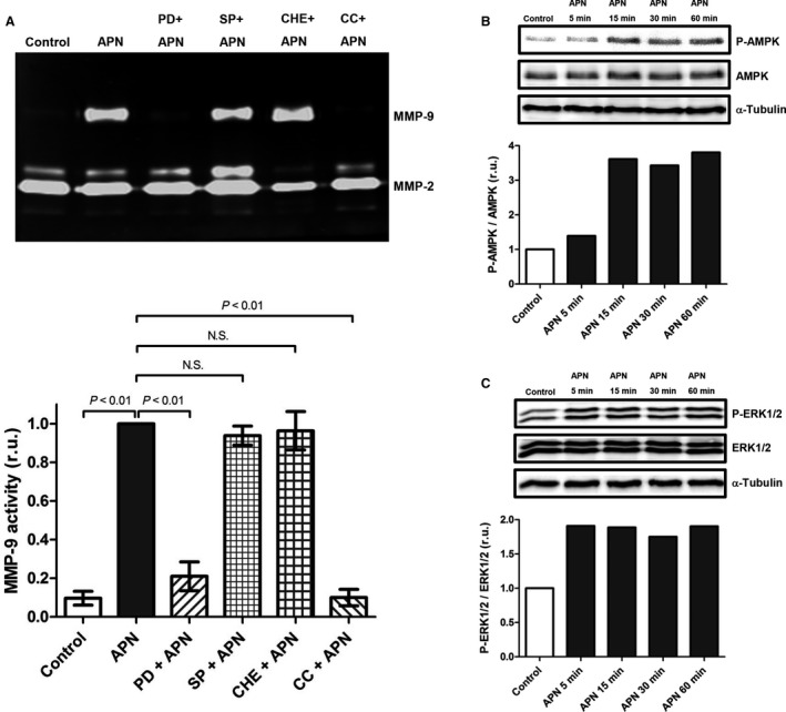
Figure 3.

APN induces up‐regulation of MMP‐9 protein expression in cardiac fibroblasts via activation of AMPK and ERK1/2. (A) Cardiac fibroblasts were pretreated with ERK1/2 inhibitor PD 098059 (PD, 20 μmol/L), JNK inhibitor SP 600125 (SP, 20 μmol/L), PKC inhibitor Cherylerythrine (Che, 5 μmol/L), and AMPK inhibitor Compound C (CC, 20 μmol/L) for 1 h followed by stimulation with APN (20 μg/mL) for 24 h. Upper panel: MMP activities in conditioned media were measured by gelatin zymography. Lower panel: Bar graph indicating quantified gelantinolytic MMP‐9 activities. Results are presented as Mean ± SEM in relative units (n = 6 independent experiments). Statistical differences were assessed using the Kruskal–Wallis test followed by post hoc testing via Mann–Whitney U test for pair‐wise comparisons between individual groups. (B) Cardiac fibroblasts were incubated with APN (20 μg/mL) for time periods of 5–60 min. Phosphorylation status of AMPK was determined by immunoblot. Quantification of α‐Tubulin expression was used as loading control. (C) Cardiac fibroblasts were incubated with APN (20 μg/mL) for time periods of 5–60 min. Phosphorylation status of ERK1/2 was determined by immunoblot. Quantification of α‐Tubulin expression was used as loading control.
