Figure 1.

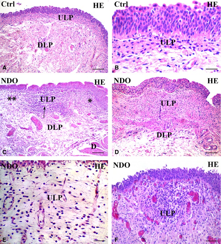
Upper lamina propria (ULP). H&E. (A, B) controls. Several rows of cells with an oval body and long, thin processes oriented parallel to the urothelium are stratified and form a network with narrow meshes. (C–F) NDO patients. The ULP is thicker than in controls: compare A with C and D. In (C) alternation of small areas where the cells are stratified and, as in controls, form a network with narrow meshes (asterisk) and areas where the stratification is less evident and the network has large meshes (double asterisks); in between a large group of immune cells (arrow). (D) non‐responder patient. The ULP is thicker than in controls and in all other patients (compare with A and C) and the cellular rows are close to each other leaving a narrow space in between. (E) the ULP network has large meshes containing a cell infiltrate and a clear (oedematous) extracellular matrix. The thin TC processes clearly make a network by contacting with each other. (F) the cell infiltrate is organized in large groups of immune cells, most of which are plasma cells, surrounded by numerous capillaries. Numerous and hyperemic capillaries are also visible under the urothelium. DLP: deep lamina propria; D: detrusor. Calibration bar: A, C, D = 100 μm; B, E = 25 μm; F = 50 μm.
