Figure 3.

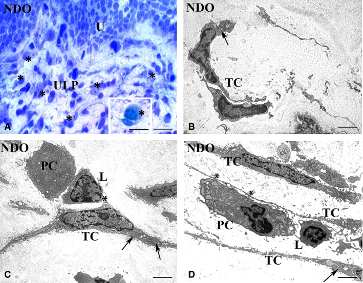
Upper lamina propria (ULP). Typical TC, NDO patients. (A) semithin section, toluidine blue stained. An area of the TC network made by regularly stratified cells. Many of these TC have an oval body and long and thin processes. Numerous immune cells (asterisks), many of which are close to the TC, are present. Inset in A: Detail of one plasma cell (asterisk) contacting a TC. (B–D) transmission electron microscopy. The cells identifiable as typical TC for their small oval body and long and thin processes share numerous RER cisternae in the body and processes (arrows). These TC are often near or make contacts (asterisks) (C, D) with the immune cells. U: urothelium; PC: plasma cell; L: lymphocyte; TC: telocyte. Calibration bar: A = 25 μm; Inset A = 10 μm; B–D = 1.6 μm.
