Figure 8.

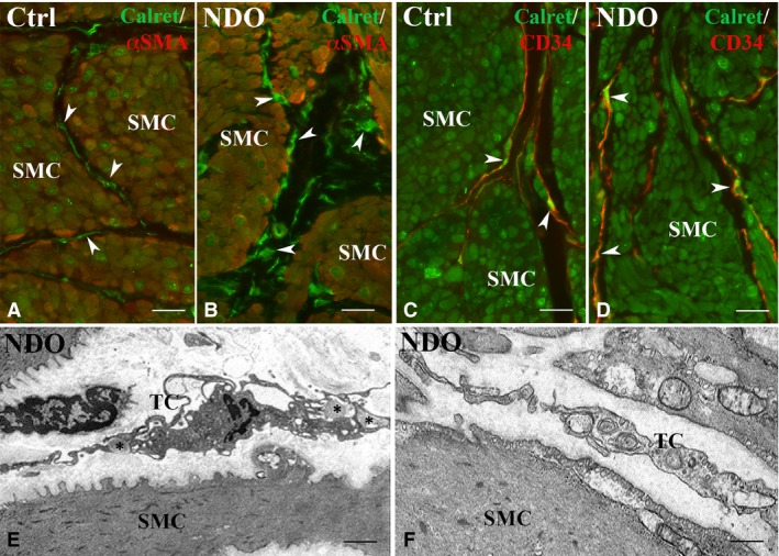
Detrusor. Immunohistochemistry. (A, B) Calret (green) and αSMA (red) labelling. In controls (A) and NDO patients (B) the smooth muscle cells express both markers while the TC (arrowheads) express only Calret. (C, D) Calret (green) and CD34 (red) labelling. In controls (C) and NDO patients (D), the smooth muscle cells express only Calret while TC express both markers (arrowheads). In NDO patients, the cells identifiable as TC are richer in Calret. Transmission electron microscopy. (E, F) NDO patients. The TC are rich in RER cisternae and caveolae. In (E), the TC processes embrace bundles of collagen fibrils (asterisks). SMC: smooth muscle cells. Calibration bar: A–D = 25 μm; E = 1.6 μm; F = 0.5 μm.
