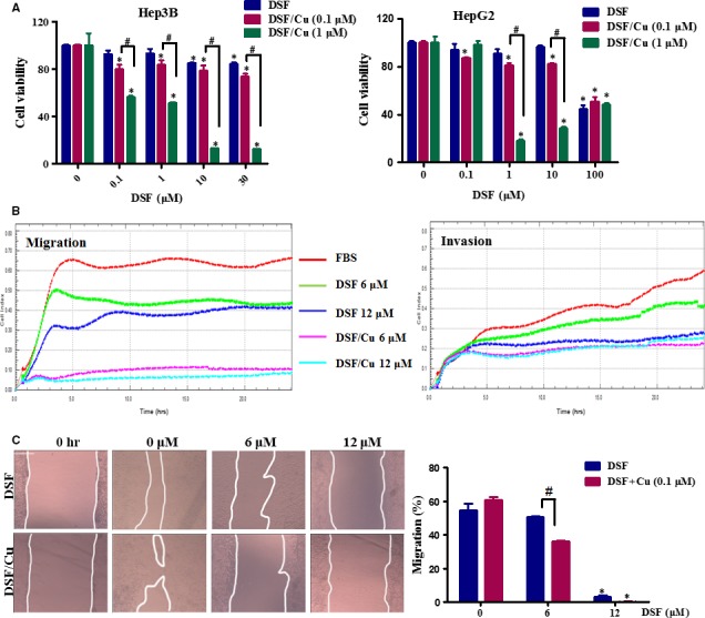
Figure 1.

DSF/Cu inhibits the migration of Hep3B cells. (A) Effect of DSF/Cu on the viability of hepatic carcinoma cells. Hep3B and HepG2 cells were incubated with increasing concentrations of DSF with or without Cu (1 and 0.1 μM) for 24 hrs. Cell viability was determined by MTT colorimetric assay. The percentage of viable cells was calculated by comparison to the absorption of control cells (0.1% DMSO), which was set as 100%. The data are reported as mean ± S.E.M. of three independent experiments. *P < 0.05, significantly different compared with the DMSO‐treated control. # P < 0.05, versus the indicated groups. Data were compared by one‐way anova followed by Dunnett's test. (B) A Real‐time measurement of the migration and invasion of Hep3B cells during 24 hrs treatment with DSF (6, 12 μM) with or without Cu (0.1 μM). (C) Scratch‐wound healing recovery assay following a 24‐hrs exposure of Hep3B cells to the indicated concentrations of DSF/Cu. The wound area was used to quantify the extent of wound healing in each group. The values obtained are expressed as a migration percentage, setting the gap area at 0 hr as 0%. Scale bar, 40 μm. *P < 0.05, significantly different compared with the control group; # P < 0.05 versus the indicated groups. Comparisons were made by one‐way anova followed by Dunnett's test. The photographs were taken at the magnification of ×100.
