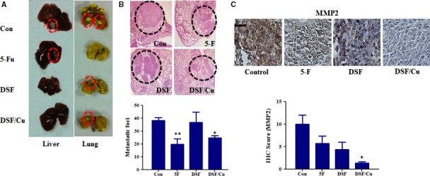
Figure 6.

In vivo anti‐metastatic activities of DSF/Cu in mice subcutaneously implanted with Hep3B cells. Mice with subcutaneous xenografts of human Hep3B cells were randomly divided into four groups (n = 8 per group) and given injections of saline (vehicle control), 5‐Fu or DSF (60 mg/kg, i.v.) with or without Cu (0.96 mg/kg, i.g.) twice a week for 29 days. The tumours were then removed for analysis. (A) Liver and lung metastasis was observed. Only a few metastases (red dashed circles) were found in mice treated with DSF/Cu. (B) Representative haematoxylin and eosin (HE) staining confirmed the development of tumours (black dashed circles) in lung tissue. The lower panel shows the number of metastatic nodules that were counted on the lung surface. (C) Immunohistochemical staining and quantification of MMP2. Scale bar, 20 μm. *P < 0.05, significantly different compared with the vehicle‐only control group; **P < 0.01 compared with control group.
