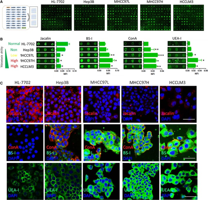
Figure 1.

Identification of metastasis‐associated lectin binding to metastatic hepatoma cells. (A) Representative lectin microarray binding patterns of the normal liver cell HL7702 and four HCC cells with difference in metastatic capacity. (B) Four lectins exhibited different extents of binding to the normal liver cell HL7702 and four HCC cells with difference in metastatic capacity. The indicated intensities are represented as the median values ± standard deviation (S.D.). (C) Incubation of Cy3‐ or Cy5‐conjugated Jacalin, ConA, BS‐I and UEA‐I and direct inspection with microscopy further confirmed the cell‐binding tendencies of these lectins, Bar = 50 μm.
