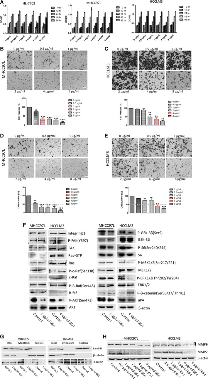
Figure 2.

Lectin BS‐I inhibited migration and invasion of HCC cell by suppressing AKT/GSK‐3β/β‐catenin pathway. (A) Cell viabilities of normal liver cell HL7702 and four HCC cells with difference in metastatic capacity treated with BS‐I at different concentrations. (B) Migration assay for MHCC97L cells treated with BS‐I at different concentrations. Data represent the means ± S.D. from three repeated experiments, *and*** represent P < 0.05 and P < 0.0001, respectively. (C) Migration assay for HCCLM3 cells treated with BS‐I at different concentrations. Data represent the means ± S.D. from three repeated experiments, ** represents P < 0.001. (D) Invasion assay for MHCC97L cells treated with BS‐I at different concentrations with collagen pre‐coated inserts. Data represent the means ± S.D. from three repeated experiments, **and*** represent P < 0.001 and P < 0.0001, respectively. (E) Invasion assay for HCCLM3 cells treated with BS‐I at different concentrations with collagen pre‐coated inserts. Data represent the means ± S.D. from three repeated experiments, **and*** represent P < 0.001 and P < 0.0001, respectively. (F) Western blot detected the effects of 1 μg/ml and 4 μg/ml BS‐I on the expression of related molecules of RAS/RAF/MEK/ERK, integrin/FAK and AKT/GSK‐3β/β‐catenin pathways in MHCC97L and HCCLM3 cells, respectively. (G) Western blot detected the effects of 1 μg/ml and 4 μg/ml BS‐I on β‐catenin expression. (H) Western blot detected MMP2 and MMP9 expression in MHCC97L and HCCLM3 cells treated with BS‐I at different concentrations.
