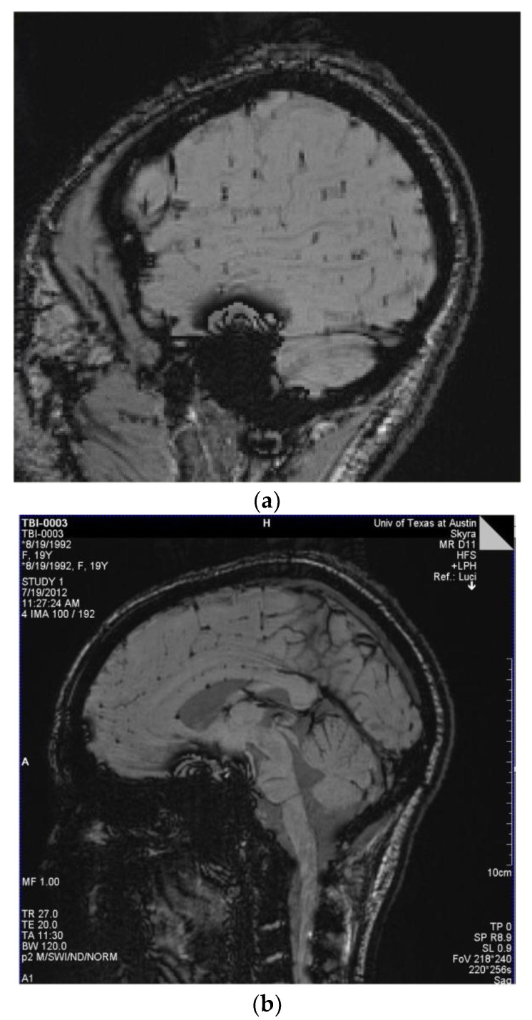
Figure 3.
(a) Players 29 and 16: Susceptibility-weighted imaging (SWI) demonstrating multiple small areas of signal loss in the base of the sulci. Paramagnetic compounds including deoxyhemoglobin, ferritin, and hemosiderin from the hemorrhages distort the magnetic field resulting in the signal loss. These images are consistent with a primary site of vascular injury and bleed into the brain parenchyma at the base of the sulci; (b) SWI Image of Player 12 demonstrating multiple small areas of signal loss in the base of the sulci. Paramagnetic compounds including deoxyhemoglobin, ferritin and hemosiderin from the hemorrhages distort the magnetic field resulting in the signal loss.

