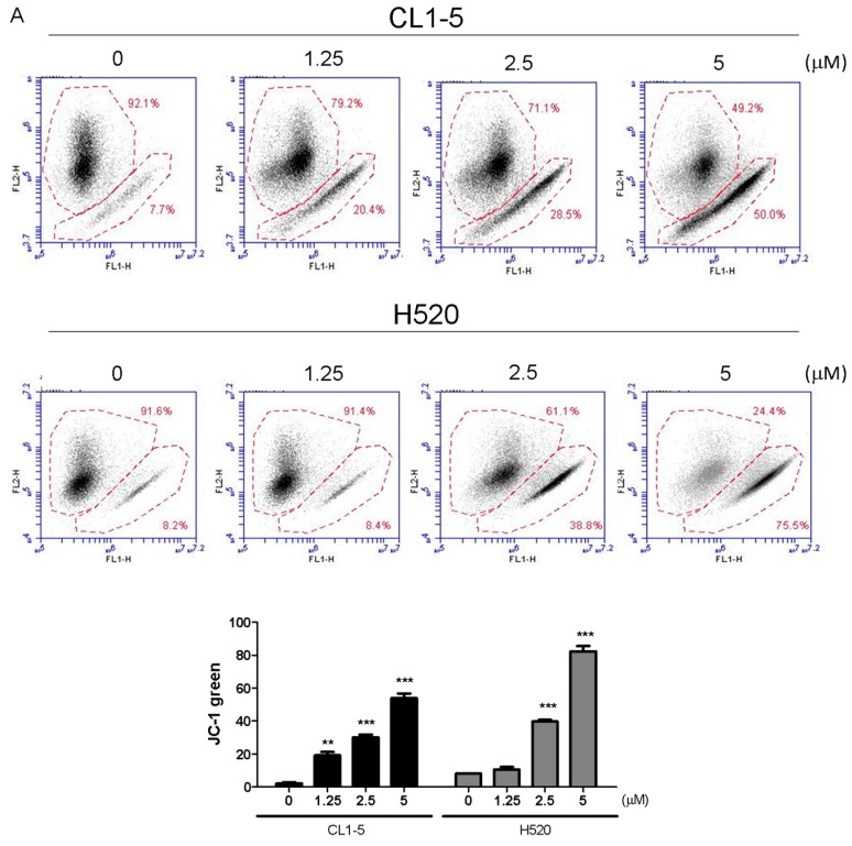
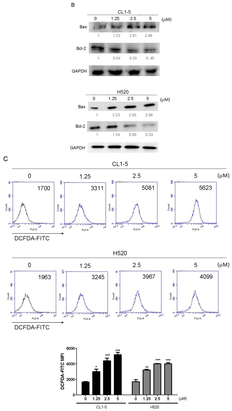
Figure 4.
A 24 h incubation with Lobocrassin B at concentrations of 2.5 and 5 μM caused mitochondrial dysfunction and ROS accumulation in CL1-5 and H520 cells. (A) The dissipation of ΔΨm was observed detecting by stained with JC-1 dye and analyzed by flow cytometry. (B) Lobocrassin B enhanced the levels of Bax and reduced the expression of Bcl-2. The fold of band intensity compared to related controls was marked respectively. (C) Lobocrassin B also increased intracellular reactive oxygen species (ROS) levels by stained with DCFH-DA dye and analyzed by flow cytometry. The bar data shown represent the mean ± SD of samples from three wells. * p < 0.05; ** p < 0.01; *** p < 0.001 compared to control group (One-Way ANOVA). All data are representative of at least three independent experiments.


