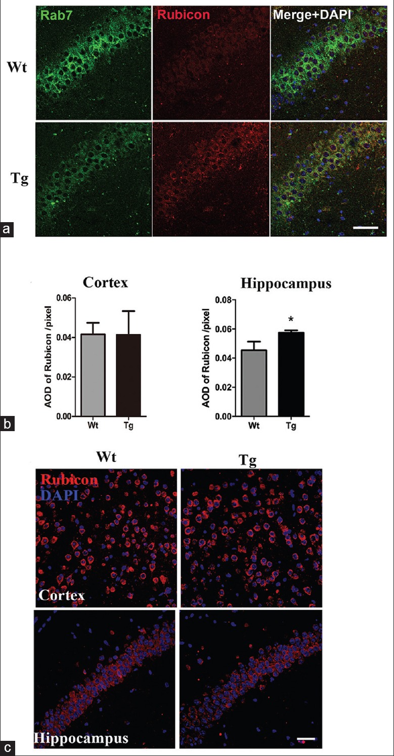
Figure 3.

Rubicon in 7- and 12-month-old APP/PS1 mice. (a) Double-labeled immunofluorescence detection of Rubicon (red) and Rab7 (green) in 12-month-old Wt and Tg hippocampus (n = 4). Scale bar = 50 μm. (b) Quantitative analysis of AOD of Rubicon in 12-month-old APP/PS1 cortex and hippocampus. *P < 0.01. (c) Immunofluorescence of Rubicon (red) in 7-month-old cortex and hippocampus of Wt and Tg mice (n = 4). Scale bar = 40 μm. AOD: Average optical density; Wt: Wild type; Tg: Transgenic.
