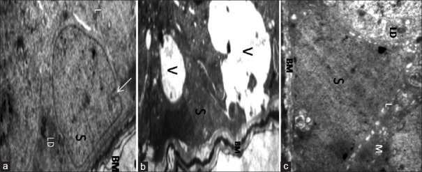
Figure 1.
Transmission electron micrograph images of Sertoli cells. (a) Control group. Sertoli cell (S) with regular euchromatic nuclei resting on a regular BM. Also LD, lysosomes (l) can be seen (original magnification ×6000). (b) Permethrin group. Sertoli cell with irregular nucleus (S) and BM. There are large vacuoles (V) in the cytoplasm (original magnification ×6000). (c) Permethrin-onion group. Sertoli cell with regular euchromatic nuclei and prominent nucleolus (S), decreased number of vacuoles compared with Permethrin group. LD (original magnification ×6000). BM: Basement membrane; LD: Lipid droplets.

