Figure 1.
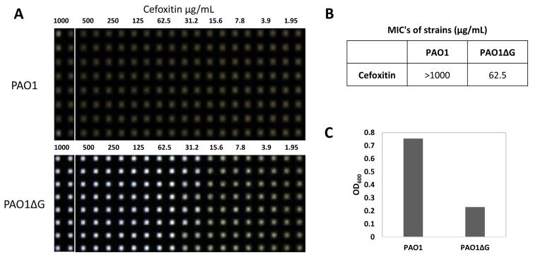
Confirmation of PAO1 pharmacology in 1536wpf. 0.5 E6 CFU/mL bacteria cultured for 17h at 37C in CAMHB exposed to 2 fold dilutions of Cefoxitin antibiotic (A). Digital images acquired on Brooke’s Plate Auditor (aka HIAPI) representing bacterial growth (brown wells) and death (clear wells). Representative Cefoxitin MIC of PAO1 vs PAO1ΔG. Each dilution encompasses two columns (B). Deletion of the AmpG gene renders PAO1 susceptible to Cefoxitin (MIC 62.5ug/mL; n=16 replicates/concentration). Absorbance measurements graphed as OD600 vs PAO1 and PAO1ΔG in the presence of 62.5ug/mL Cefoxitin – S:B 3.3, Z′ 0.88 (C).

