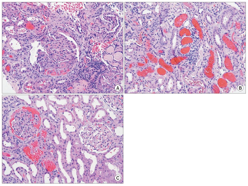
Figure 2. Light microscopy kidney biopsy findings (H&E stain).
(A) A single small segmental cellular crescent is noted in one out of 68 glomeruli with open capillary loops (×100). (B) Numerous occlusive red blood cell casts were seen in the tubules. Acute tubular necrosis is present (×100). (C) Red blood cells were filling Bowman’s space in some glomeruli (×200).

