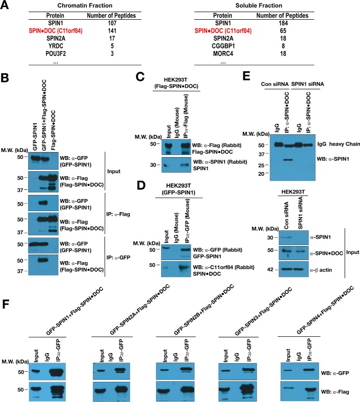
Figure 1.
SPIN1 interacts with SPIN·DOC in cells. A, lists of SPIN1-associated proteins identified in both the chromatin and soluble fraction by mass spectrometry analysis. B, GFP-SPIN1 and FLAG-SPIN·DOC was transiently transfected alone or together into HEK293T cells. Cell lysates were immunoprecipitated with α-FLAG or α-GFP and Western-blotted (WB) with the indicated antibodies. MW, molecular weight. C, HEK293T cells were transiently transfected with FLAG-SPIN·DOC, and whole-cell lysates were immunoprecipitated with α-FLAG or IgG control antibodies. Western blot analysis was performed with α-FLAG and α-SPIN1 antibodies. D, HEK293T cells were transiently transfected with GFP-SPIN, and whole-cell lysates were immunoprecipitated with α-GFP or IgG control antibodies. Western blot analysis was performed with α-FLAG and α-SPIN·DOC antibodies. E, HEK293T cells were subjected to knockdown with SPIN1 siRNA (or control (Con) siRNA) for 24 h, and then the whole-cell lysates were subjected to immunoprecipitation with α-SPIN·DOC or IgG control antibodies. Western blot analysis was performed using α-SPIN1. F, HEK293T cells were co-transfected with FLAG-SPIN·DOC and GFP-SPIN family members (SPIN1, 2A, 2B, 3, and 4), respectively. Whole-cell lysates were immunoprecipitated with α-GFP or IgG control antibodies and blotted with the indicated antibodies. In all depicted experiments, the input represent 2% of the sample used in the co-IP.

