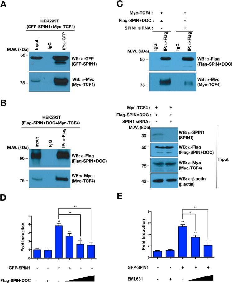
Figure 4.
SPIN·DOC blocks SPIN1-mediated Wnt/TCF-4 signaling. A and B, GFP-SPIN1 and Myc-TCF4 (A) and Myc-TCF4 and FLAG-SPIN·DOC (B) were transiently co-transfected into HEK293T cells. The whole-cell lysates were immunoprecipitated and blotted with the indicated antibodies. MW, molecular weight; WB, Western blot. C, HEK293T cells were transfected with SPIN1 siRNA or control siRNA. The whole-cell lysates were immunoprecipitated with a-FLAG or IgG control antibodies, and then the indicated proteins were identified by Western blot analysis. D, Wnt-responsive luciferase reporter assays were performed in transfected T778 cells in the presence of GFP-SPIN1 and increasing amounts of FLAG-SPIN·DOC plasmid DNA. E, Wnt-responsive luciferase reporter assays were performed with GFP-SPIN1–transfected T778 cells that were incubated with or without EML631 (15, 30 mm). Error bars show S.D. *, p < 0.05; **, p < 0.01; ***, p < 0.001; two-tailed Student's t test. The input represents 2% of the lysate that was used in every experiment.

