Figure 2.
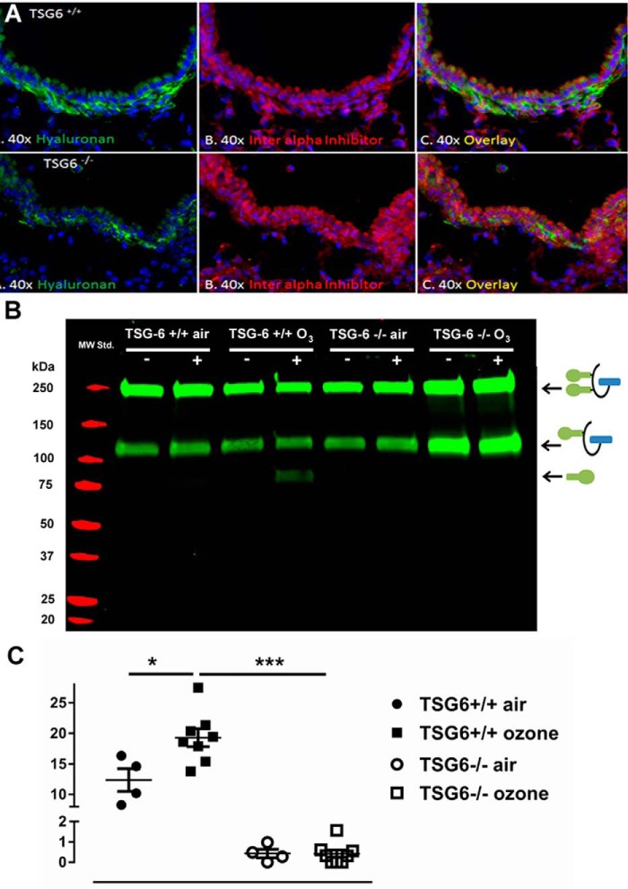
HA–HC deposition after ozone exposure. Mice were exposed to 2 ppm ozone for 3 h and sacrificed 24 h later. A, lung tissue was fixed in paraffin and stained for hyaluronan and IαIp. In TSG-6–sufficient mice, there is immunohistochemical evidence of HA–HC deposition in the subepithelial space, which is absent in TSG-6–deficient mice. B, tissue was digested with hyaluronidase, and released heavy chains were detected by Western blotting. Detection of HA-bound HC shows an increase of HA–HC complex formation only in TSG-6–sufficient mice after ozone exposure (note free HC band at ∼80 kDa, which appears only after hyaluronidase treatment). Schematics of the full IαI molecule (at 250 kDa), the pre-αI molecule (at 120 kDa), and the free HC (at 80 kDa) are provided to the right of the blot for clarification. +, hyaluronidase treatment; −, no hyaluronidase treatment. C, quantification of HA-bound HC shows a significant increase in TSG-6–sufficient mice after ozone exposure. TSG-6–deficient mice have virtually no HA-bound HC before or after ozone exposure. Results are represented as means ± S.E. (error bars). The experiment was repeated at least twice. n = 4 (air exposure) to 8 (ozone exposure). *, p < 0.05; ***, p < 0.001, ANOVA with Tukey's multiple-comparison correction.

