Figure 7.
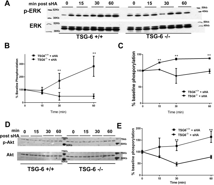
ERK and Akt activation after sHA exposure is TSG-6–dependent. Primary mouse ASMC were cultured to 80% confluence and then washed repeatedly and serum-starved for 4 h and finally exposed to sHA or vehicle, as indicated. A, Western blotting for phospho-p42 and phospho-p44 ERK. B, quantification of phospho-p42 by densitometry shows a rapid increase of phosphorylated protein in TSG-6–sufficient cells, whereas deficient cells show no change. C, quantification of phospho-p44 by densitometry shows a rapid increase of phosphorylated protein in TSG-6–sufficient cells, whereas deficient cells show no change. D, Western blot for phospho-Akt at threonine 308 shows increased protein phosphorylation after sHA exposure, which is absent in TSG-6–deficient cells. E, quantification shows that the increase in phosphorylation is significant in TSG-6–sufficient cells. Results are represented as means ± S.E. (error bars). The experiment was repeated at least three times. n = 3–6/condition. **, p < 0.01, TSG-6–sufficient versus TSG-6–deficient, ANOVA with Tukey's multiple-comparison correction.

